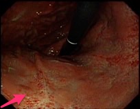
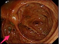
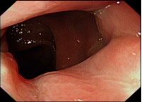
After admission, paracentesis was performed, and the ascites showed WBC 3520/cmm, lymphocyte 54 % , albumin 3.0 g/dL and AFS(-). SAAG is 1.1(serum albumin 4.1 g/dL). Cytology was negative. Abdomen CT showed peritoneal thickening (omental caking) and one heterogenous soft tissue at pelvic area (doughy abdomen). Due to the possibility of carcinomatosis of unknown origin, we arranged EGD and colonoscopy that showed gastric ulcers at antrum and lower body and diverticula at ascending and sigmoid colon. However, gastric biopsies showed chronic gastritis with intestinal metaplasia and no evidence of H. pylori infection. Laparoscopy showed peritoneum studded with multiple whitish nodules and so-called "Violin-string" fibrinous strands. Peritoneal biopsies showed granulomatous inflammation. We also arranged thoracentesis that demonstrated exudative effusion. Acid-fast stains of ascites and peritoneal biopsy were negative. However, tuberculous peritonitis still could not be ruled out. Hence, we started HERZ therapy. Culture grew Mycobacterium tuberculosis one month later.
Tuberculous peritonitis

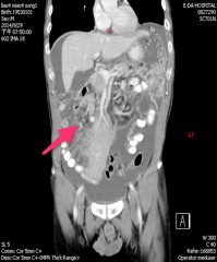
Suspect peritoneal carcinomatosis with omental cake

義大醫院‧總機電話:07-615-0011‧07-952-0011 地址:82445高雄市燕巢區角宿里義大路1號
語音‧人工掛號專線:07-615-0900‧07-615-0911 週一至週五 07:30~16:30 週六 07:30~11:30
© Copyright 2014. "EDAGI" All rights reserved.