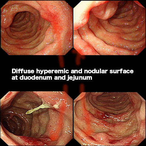
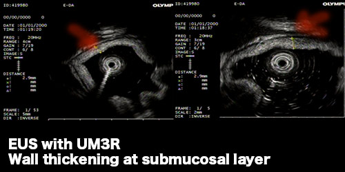
Follow-up EGD showed that mucosa of bulb and second portion was fragile and easily bleeding. Several polypoid lesion with ulcers were noted at post bulbar area s/p biopsy. After EGD, we changed to side-view endoscope. The papilla was invisible. Elevated lesion with fragile mucosa was noted at the ampular portion s/p biopsy.
The section shows duodenal mucosa with deposits of hyalinized material in lamina propria and mucosa muscularis.
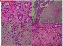
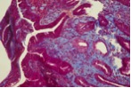
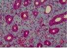
This case is consulted. Professor Shun in NTUH think this hyalinized material is amyloidosis as evidenced by positive Congo red stain. The case is unlike collagenous duodenitis, usually deposite in basement memberane area. So he suggestd to check the rectum or oral mucosa to rule out primary amyloidosis.
Oral ulcer(-), arthritis(-),rash(-),photosensitivity(-),leg edema(-),Raynaud phenomenon(-),dry month or eye(-),back stiffness(-)
Intestine amyloidosis
義大醫院‧總機電話:07-615-0011‧07-952-0011 地址:82445高雄市燕巢區角宿里義大路1號
語音‧人工掛號專線:07-615-0900‧07-615-0911 週一至週五 07:30~16:30 週六 07:30~11:30
© Copyright 2014. "EDAGI" All rights reserved.